
안녕하세요.
Dr.A 입니다.
오늘은 무릎 초음파를 볼때 확인해야하는 간단한 구조물들 대해 알아보도록 하겠습니다.
Anterior scan
1) 환자를 supine position 으로 자세를 취한 뒤 knee를 최대한 flexion 시킨다.

2) Probe를 longitudinal하게 부착하여 femur, tibia 위쪽의 patella를 관찰할 수 있고 해당 부위 위쪽으로 지나가는 quadriceps tendon과 tibial tendon을 확인할 수 있는데 hyperechoic한 fibrillar pattern으로 나타나게 된다. 이때 patella tendon은 tibial tuberosity까지 부착하는 모습을 확인할 수 있다.
3) knee extensor로 작용하는 quadriceps tendon과 tibial tendon을 확인하기 위해서 tendon tightening을 통해 잘 볼 수 있도록 한다.
4) patella 위쪽으로 surapatella fat pad와 더 위쪽으로는 분리되어있는 prefemoral fat pad를 확인할 수 있다.
1- suprapatella fat pad 2- prefemoral fat pad
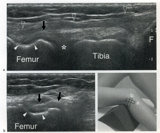
5) Transverse view로 부착하는 경우 knee를 최대한 flexion 시킬때 trochlear cartilage의 노출을 확인할 수 있으며 medial facet과 lateral facet의 superficial하게 존재하는 모습을 확인할 수 있다.
Medial scan
1) 환자를 검사측으로 돌려 눕힌 다음 knee를 60' 정도 flexion 시킨다.
2) Femur의 medial epicondyle과 tibial의 medial condyle의 prominent한 부분을 촉지하여 probe를 부착하는 경우 아래에 지나가는 medial collateral ligament complex를 확인할 수 있다.
3) Medial collateral ligament는 superficial layer과 deep layer로 나누어지게 되는데,
superficial layer는 thick하고 straight, fibrillar pattern으로 나타나며 deep layer는 hyperechoic thin band로 나타난다.
Lateral scan
1) 환자를 검사측 반대편으로 돌리고 knee를 30'가량 flexion 시킨다.
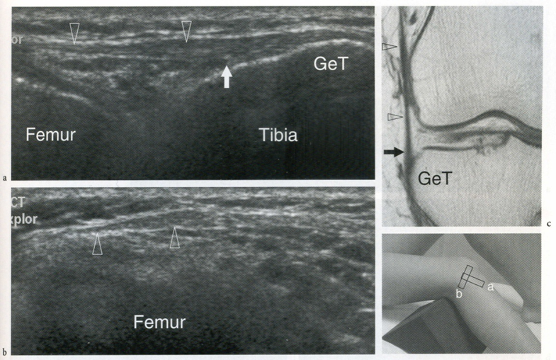
2) 이때 femur의 lateral epicondyle, Gerdy's turbercle, fibular head 세가지 landmark 구조물을 촉지한다.
3) Lateral epicondyle과 Gerdy's turbercle을 연결하는 위치로 probe를 부착하게되면 iliotibial tract를 확인할 수 있다. 아래 그림처럼 Gerdy's turbercle과 femur lateral epicondyle이 hyperechoic band 형태로 나타난다.
4) Lateral femur epicondyle과 fibular head 사이로 probe를 옮기게 되는 경우 Lateral collateral ligament를 확인할 수 있다.
5) 이때 lateral collateral ligament의 deep portion에서 popliteal tendon의 transverse view를 관찰할 수 있다.
'We need to talk about rehab > Sonography' 카테고리의 다른 글
| Ankle joint/발목 초음파 보는법 (0) | 2021.10.01 |
|---|---|
| 체외 충격파 치료의 적응증. 언제 해야할까? 부작용? (0) | 2021.06.03 |